Wednesday, February 15, 2012

Infantile Hemangiomas


From Medscape Dermatology > Viewpoints

Therapy for Infantile Hemangiomas

Propanolol Blows Away Corticosteroids

Graeme M. Lipper, MD
Posted: 02/08/201


Propranolol vs Corticosteroids for Infantile Hemangiomas: A Multicenter Retrospective Analysis

Price CJ, Lattouf C, Baum B, et al
Arch Dermatol. 2011;147:1371-1376

Study Summary

Infantile hemangiomas (IH) are proliferative vascular tumors that, despite their benign nature, may cause pain, bleeding, scarring, or functional impairment with potentially life-threatening consequences. Because most IHs involute within the first decade of life, noncritical lesions are typically followed for expected gradual regression. In contrast, lesions that obstruct vision, respiration, hearing, feeding, or defecation require prompt medical or surgical intervention. Large or multifocal lesions pose significant risks, including high-output cardiac failure and cosmetic disfigurement.[1] In the past, such high-risk IHs were treated with systemic corticosteroids, an option that is limited by variable efficacy and common adverse effects such as cushingoid features, adrenal suppression, gastritis, hypertension, and growth impairment.
In 2008, French investigators made the serendipitous discovery that propranolol, a beta-blocker traditionally used for the treatment of hypertension, tachycardia, and congestive heart failure, can induce dramatic IH regression.[2]Subsequent reports confirming the impressive efficacy and safety of propranolol have been met with growing optimism.[3-5] However, one pressing question remained: How does propranolol compare with systemic corticosteroids? In a landmark multicenter study, Price and colleagues convincingly answered this question. It turns out that propranolol blows the historical gold standard out of the water.
In order to reach this conclusion, Price and colleagues retrospectively analyzed the clinical outcomes in 110 infants and children with IH (77% female; 78% located on the head and neck; mean age of treatment initiation, 4.7 years). The patients were subdivided into 2 treatment groups: those receiving propranolol (n = 68; mean treatment duration, 7.9 months; target dose, 2 mg/kg/d) and those receiving oral corticosteroids (n = 42; mean treatment duration, 5.2 months; target dose, 4 mg/kg/d). To insure comparability, investigators matched these groups for patient age and lesion size, location, and type.
Outcome measures included percentage of IH clearance (< 75% or ≥ 75%), adverse effects, percentage of patients needing subsequent surgical referral, and average treatment cost per IH. Propranolol proved superior in all of these criteria:
  1. 82% of patients treated with propranolol achieved ≥ 75% clearance vs 29% of patients treated with oral corticosteroids (P < .01).
  2. 1 of 68 patients treated with propanolol had transient hypoglycemia, 2 patients (3%) had nonspecific skin eruptions, all patients completed treatment, and no patients suffered serious adverse events, whereas all patients treated with oral corticosteroids had adverse effects, including cushingoid features (100%), gastroesophageal reflux (n = 4), and hypertension (n = 2). One patient had a life-threatening bleed due to IH ulceration eroding into the external carotid artery.
  3. 12% of patients treated with propranolol required surgery after therapy vs 29% of patients treated with oral corticosteroids.
  4. The average cost per IH treated (excluding monitoring costs and prophylaxis/treatment of adverse effects) was $205.32 for propranolol vs $416.00 for oral corticosteroids.

Viewpoint

After its recent introduction by French investigators, oral propranolol has revolutionized the management of severe infantile hemangiomas. Now, thanks to Price and colleagues' seminal study, no lingering doubt should exist that propranolol is the first-line agent for treating symptomatic or cosmetically disfiguring IH. When comparing safety and efficacy of the 2 drugs, the results are not even close.
Although "propranolol blows away corticosteroids" is the headline here, the investigators of this study also made several other important observations. For example, they found that:
  1. Propranolol induced IH regression even when treatment was initiated after the first year of IH growth, confirming previous reports that the drug works to debulk more mature lesions beyond the proliferative phase.[6]
  2. Initial treatment with oral corticosteroids followed by oral propranolol showed a trend toward slightly improved efficacy over propranolol alone, although this benefit was not statistically significant.
  3. IHs were less likely to relapse if propranolol therapy continued until at least 1 year of age.
  4. The risk for propranolol-related side effects can be minimized with proper monitoring (eg, cardiac preclearance and checking for signs or symptoms of bradycardia, hypotension, and hypoglycemia).
Finally, Price and colleagues acknowledged that at least one more critical question remains: We still do not knowhow propranolol works to slow IH and induce regression. Does it trigger endothelial cell apoptosis, inhibit proangiogenic cytokines, or cause vasoconstriction by reducing nitric oxide levels? As researchers try to solve this complex puzzle, powerful new IH treatments will surely follow
.

Thursday, February 9, 2012

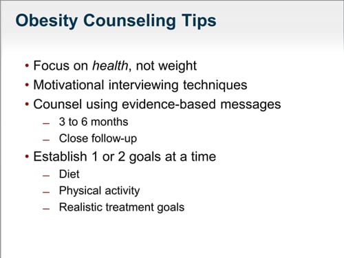
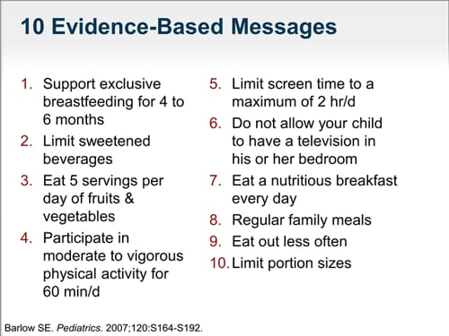
Prevent Obesity in Child

Slide 17.
Slide 16.

Chest Pain in Children - majority not cardiac


From Medscape Pediatrics > Viewpoints

Chest Pain in Children: Is It Cardiac?

William T. Basco, Jr., MD
Posted: 01/30/2012
 
 
Like this article?
Read medical news in 32 specialty
areas with our Medscape App.
Choose the specialties you want
to follow.
Learn More

Effectiveness of Screening for Life-Threatening Chest Pain in Children

Saleeb SF, Li WY, Warren SZ, Lock JE
Pediatrics. 2011;128:e1062-e1068

Chest Pain in Children

Studies have consistently shown that most chest pain experienced in childhood and adolescence is not cardiac in nature. Therefore, chest pain in children does not have the same concerning connotation that it does in adults. This study was done to determine whether unexpected cardiac events occurred in children who were evaluated in a cardiology clinic and determined to have a noncardiac origin for their pain.

Study Summary

All children were evaluated at a single medical center in Boston over a 10-year period, 2000-2009. Children were older than 6 years and had no known cardiac defects or cardiovascular disease. Children who were seen for chest pain were identified from medical records. Additional data included demographic features, clinical characteristics, cardiac testing, discharge diagnoses, other medical diagnoses, and follow-up.
Children were divided into those who had chest pain on exertion and those who had chest pain at rest. In general, children who experienced chest pain on exertion as one of the presenting symptoms had more extensive testing. ECGs were performed on all children, but additional testing varied during the study period at the discretion of the cardiologist evaluating the patient. Two sources were used to identify deaths after the visit: the National Death Index from the Centers for Disease Control and Prevention and the Social Security Death Index.
The final cohort included 3700 children. The median age at the time of the cardiac evaluation was 13.4 years, and the children had a median of 4.4 years of follow-up. Chest pain at rest was the most common scenario (56%), followed by exertional chest pain (33%). Palpitations accompanied the chest pain in 22% of patients, shortness of breath in 16%, dizziness in 11%, and syncope in 1.3%. Almost 1 of 5 children had made at least 1 emergency department visit for chest pain before the cardiac evaluation. Fifteen percent of the children had a history of asthma, and almost 2% had a history of gastroesophageal reflux. Approximately 1% had a minor congenital heart problem, and an additional 1% had an inflammatory disorder. The physical examination findings were mostly normal.
Results of cardiac evaluations. Identified cardiac abnormalities included clicks (1.4%), structural abnormalities of the sternum (1.1%), and pathologic murmurs (0.8%). The ECG evaluations were largely normal, with left ventricular hypertrophy demonstrated in only 2.5% of children and abnormal ST segments, abnormal T waves, right ventricular hypertrophy, or conduction abnormalities demonstrated in less than 1%. Echocardiography was done in 38% of patients; these evaluations were normal in 88%, and an additional 11% had incidental findings that the investigators felt were unrelated to chest pain. In 0.8% of patients, the echocardiograms identified abnormalities that might be related to the chest pain; these incidental findings included mild dilations of the aorta, mild mitral regurgitation, mitral valve prolapse, and miscellaneous problems.
Causes of chest pain. The discharge diagnoses from the cardiology clinic evaluation for chest pain were apportioned as follows:
  • Unknown origin, 52%;
  • Musculoskeletal origin, 36%;
  • Pulmonary origin, 7%;
  • Gastrointestinal origin, 3%;
  • Anxiety, 1%; and
  • Cardiac origin, 1%.
Deaths during follow-up. Among the 3700 patients, 3 deaths (0.1%) occurred during follow-up. Two of these deaths were suicides, and the third was the result of retroperitoneal hemorrhage. The researchers concluded that chest pain is a common pediatric symptom that rarely has a cardiac cause and that, in almost 18,000 patient-years of follow-up, no patient discharged from the clinic died of a cardiac condition.

Viewpoint

This is an excellent study that may or may not reassure primary care practitioners. Although the incidence of cardiac diagnoses was exceedingly low at 1%, some of the conditions that were identified are potentially serious cardiac abnormalities. Allaying residual concerns about missing even the small percentage of true cardiac diagnoses is exactly why these investigators conducted the main analyses that looked at deaths after discharge from their clinic. Children discharged with a diagnosis of chest pain are probably a selected group, so the investigators repeated the analysis to include the 41 children who had chest pain as a presenting symptom but ended up with a more severe cardiac diagnosis. This increased the overall incidence of cardiac disease in children with chest pain to 2%. Even among the 41 children with a more serious cardiac diagnosis, no deaths occurred.
It is worth noting that this is a subspecialty referral population, meaning that primary care practitioners probably filtered out many other cases of chest pain before they arrived at the pediatric cardiology clinic. Therefore, the incidence of cardiac disease is probably even lower among all children in a primary care who are experiencing chest pain. Nevertheless, Saleeb and colleagues are evaluating an outpatient application of a decision approach to referral and testing for chest pain in primary care practitioner offices, and I know that having such a validated guideline would be very welcome to most pediatric practitioners when seeing a patient with chest pain.

Friday, January 27, 2012

pediatric immunisation n anaphylaxis


From Medscape Medical News

Anaphylaxis After Pediatric Immunization Rare

Troy Brown
January 26, 2012 — A recent study found that anaphylaxis occurred rarely after pediatric immunization, and not at all after routine infant and preschool immunization.
Michel Erlewyn-Lajeunesse, MD, from the Children's Allergy Clinic, University Hospital, Southampton, United Kingdom, and colleagues described their findings in an article published online January 23 in the Archives of Disease in Childhood.
Because it is so rare, "[e]ven the largest prelicensure vaccine trials are unlikely to detect a single case, let alone provide an estimate of incidence," write the authors. "The onus for detection of anaphylaxis falls to national post-marketing surveillance...systems, all of which rely upon passive reporting of cases."
For this reason, Dr. Erlewyn-Lajeunesse and colleagues conducted a study using prospective active surveillance to determine the incidence and clinical presentation of anaphylaxis as an adverse event after immunization (AEAI) in children younger than 16 years.
The British Paediatric Surveillance Unit (BPSU) sends an "orange card" with a list of rare disorders to consultant pediatricians in the United Kingdom and Ireland each month. Physicians note the presence or absence of these disorders on the card and return it to the BPSU. Reports were collected for 13 months, from September 1, 2008, to September 30, 2009, with more than 90% of participants returning the cards (93.2% in the United Kingdom and 91.8% in Ireland).
Clinicians were instructed to report actual cases of anaphylaxis, as well as cases in which anaphylaxis was only suspected but further immunization was contraindicated. The Brighton Collaboration Case Definition (BCCD) for anaphylaxis as an AEAI was used to identify cases of anaphylaxis.
During the 13 months, 15 reports of anaphylaxis as an AEAI were made to the BPSU. Of those, 7 cases met the BCCD criteria (3 were BCCD level 1, 3 were BCCD level 2, and 1 was BCCD level 3). The child whose symptoms met level 3 diagnostic criteria was treated with intramuscular adrenaline quickly, and this may have been the reason that case was less severe.
Of the other 8 cases, 3 were withdrawn by the reporting physician, and further information was unavailable from reporting clinicians for 3 others. One child experienced a hypotonic hyporesponsive episode, and the last case was not included because it occurred outside of the study period.
In 6 of the 7 cases, the reporter believed the episode was caused by the immunization, and in the other case, the reporter believed it was probably related.
All but 1 of the children were receiving the vaccination for the first time. One child had received the immunization before without incident, and 2 of the children were given several immunizations at the same appointment.
The study analyzed reports related to 3 human papilloma virus vaccines (Cervarix, GlaxoSmithKline), 2 single-component measles vaccines (Rouvax, Sanofi Pasteur MSD), a meningococcal C conjugate vaccine, a school leaver's booster (probably tetanus/inactivated polio virus), an inactivated typhoid vaccine (Typhim Vi, Sanofi Pasteur MSD), a quadrivalent meningococcal polysaccharide vaccine (ACWY Vax, GlaxoSmithKline), and a hepatitis A vaccine (Havrix Junior Monodose, GlaxoSmithKline).
Symptoms began within 15 minutes of administration in 3 children, and 30 minutes or later in the other 4 children. One of those 4 children experienced symptoms 120 minutes after immunization.
Intramuscular adrenaline was administered to 6 children, intravenous fluids were given to 3 children, and 1 child received salbutamol nebulization. Corticosteroids were given to 2 children, H1 histamine receptor inverse agonists were given to 5 children, and 1 child improved without treatment.
No child required pediatric intensive care admission, but 3 required emergency department treatment, 4 needed acute pediatric treatment, and 2 received pediatric outpatient care after the occurrence. All children recovered completely with no sequelae.
Preexisting atopic disease made it necessary for 3 children to carry injectable adrenaline: 1 child had multiple food allergies, and 2 had idiopathic urticaria with anaphylaxis.
During the study period, 16,625 doses of single-component measles vaccine were ordered by United Kingdom and Ireland. There were 2 reports of anaphylaxis AEAI, amounting to an incidence of 12 cases per every 100,000 immunizations.
Of 2,081,272 vaccines given during the study period to children aged from 12 to 19 years (most younger than 16 years), there were 3 events, amounting to an incidence of 1.4 cases per million immunizations.
"The important thing for clinicians to remember is that vaccines are extremely safe," Vivian Hernandez-Trujillo, MD, director of the Division of Allergy and Immunology at Miami Children's Hospital in Florida, told Medscape Medical News in a telephone interview.
"Adverse reactions to vaccines may occur, [but] anaphylaxis is extraordinarily rare," she added.
"No events were related to routine infant and preschool immunizations despite over 5.5 million primary schedule vaccines being delivered in this time period. Some children had delayed onset of symptoms and this should be considered in those at higher risk of anaphylaxis," the authors conclude.
Arch Dis Child. Published online January 23, 2012. Abstract

Wednesday, January 25, 2012

Clarifying Meningococcal Booster Dose Recommendations

From CDC Expert Commentary Amanda Cohn, MD 01/17/2012 Hi, I am Dr. Amanda Cohn, a pediatrician and epidemiologist. Thanks for tuning in to this CDC Expert Video Commentary on Medscape. In January 2011, recommendations were made by the Advisory Committee on Immunization practices (ACIP) for adolescents to receive a booster dose of meningococcal conjugate vaccine. Today I am going to explain the rationale behind this recommendation and answer frequently asked questions about implementation. First, some background. When this vaccine was first recommended for adolescents in 2005, the expectation was that protection would last for 10 years. However, currently available data suggest that immunity wanes much earlier than 10 years. In fact, only about half of adolescents are still protected 5 years after administration of the initial dose. Based on that information, a single dose for all 11- to 18-year-olds, recommended to be given at age 11-12 years, may not offer continued protection through the period when risk for meningococcal infection is highest, 16-21 years of age. Therefore, ACIP voted to recommend a booster dose of meningococcal conjugate vaccine for adolescents. This booster dose is recommended to be given at 16 years of age, following the routine first dose at age 11-12 years. Unfortunately, not all adolescents receive these doses on time, so here are some key points about when to give the booster dose. For adolescents who receive the first dose at age 13-15 years, a 1-time booster dose should be administered, preferably between the ages of 16 and 18 years. There is no need to wait 5 years from the first dose before administering the booster. Rather, give the booster dose any time after the child's 16th birthday. Eight weeks is the minimum interval between doses. If the teen is younger than 16 years of age, but the clinician believes this is a situation where there may not be another opportunity to provide the booster dose, the second dose can be given prior to 16 years of age. The booster dose is not recommended for adolescents who receive their first dose of meningococcal conjugate vaccine after their 16th birthday. What about college requirements? Meningococcal vaccination is required to attend many colleges, but which kids going off to college still need the booster dose? You may have a patient going off to college who requires vaccination. Many colleges will consider any dose given within 5 years prior to matriculation as valid. However, the recommendation for providers is to still follow the new guidelines: Administer a dose (a booster dose or the first dose if the adolescent is unvaccinated) after the 16th birthday and prior to college. Ideally, we want kids to get the booster dose before they reach the age when they are at greatest risk. All college freshmen living in a dormitory are recommended to be fully vaccinated. The booster dose may still be given to college students not living in dorms but is not routinely recommended. Which vaccine should be used? Only the meningococcal conjugate vaccine is recommended for adolescents. However, if the first dose of meningococcal vaccine was administered as polysaccharide vaccine, it is still counted as valid in the adolescent schedule. The booster dose of meningococcal vaccine for adolescents should always be a conjugate vaccine. The 2 licensed products, Menactra® and Menveo®, are interchangeable. If polysaccharide vaccine is inadvertently administered as the booster dose, revaccination with conjugate vaccine is recommended 8 weeks later. We want to protect as many kids as possible from this rare but often devastating disease. For more information on meningococcal disease and vaccine recommendations, visit www.cdc.gov/meningococcal. Thanks for tuning in today.

HPV & Sex

From Medscape Infectious Diseases HPV & Sexual activity - any relationship? Paul A. Offit, MD 01/17/2012 Hi, my name is Paul Offit. I am talking to you today from the Vaccine Education Center at the Children's Hospital of Philadelphia. What I want to talk about is an article on the human papillomavirus (HPV) vaccine that recently appeared in American Journal of Preventive Medicine. The first HPV vaccine, called Gardasil®, contains serotypes 6, 11, 16, and 18 and came out in 2006. At that time, it was recommended for girls only. This past year, the vaccine was also recommended for boys. When the vaccine came out, there were a number of issues that were of concern to parents. People wondered whether the vaccine was really safe. There were questions about whether it caused blood clots, with consequent strokes and heart attacks. There were questions about whether it caused chronic fatigue syndrome. More recently, in September 2011, during the Republican national debates, Michele Bachmann raised the question of whether the HPV vaccine could cause severe developmental delays, which she referred to as "mental retardation." So, there has been a lot of fear surrounding this vaccine. In fact, none of those concerns are true. A number of studies have shown that the HPV vaccine does not cause chronic fatigue syndrome, blood clots, strokes, or heart attacks. But another issue was raised that really hasn't been addressed until this article was published, and that is the question of whether getting an HPV vaccine increases your desire for sexual activity -- or, said another way, promiscuity. The researchers at the Centers for Disease Control and Prevention, headed by Lauri Markowitz, looked at this. In their article titled "Human Papillomavirus Vaccine and Sexual Behavior Among Adolescent and Young Women," they looked at whether those who got the HPV vaccine were more likely to be promiscuous than those who did not. The answer was, not surprisingly, no. It doesn't make sense that that would have ever been true. First of all, no vaccine is 100% effective. Second, this particular vaccine protects against about 70% of strains that cause cervical cancer and 90% of strains that cause anal and genital warts. So, it's not even 100% effective against all strains of HPV. Obviously, the vaccine does not prevent other sexually transmitted diseases, such as syphilis, gonorrhea, chlamydia, or herpes. The concern never made sense. It's like making an argument that once I get a tetanus-containing vaccine, I can feel comfortable running through a bed of rusty nails. I don't think that is true either. I think we can now feel comfortable about the safety of this vaccine and also the notion of whether it increases sexual activity or promiscuity. The problem with HPV vaccine is that people haven't been very good about getting it. Only about one third of girls and young women for whom this vaccine is recommended get it. Hopefully, we can be better at encouraging vaccine use, and hopefully this study will make people feel more comfortable about the vaccine. Thank you. Related Resource Prevent HPV

Hair Loss and its Management in Children

From Expert Review of Dermatology Vibhu Mendiratta; Masarat Jabeen 01/15/2012; Expert Rev Dermatol. 2011;6(6):581-590. © 2011 Expert Reviews Ltd. Abstract Hair loss in children can cause psychological stress to the parent and patient alike. Alopecia can be classified into congenital and acquired. Commonly encountered causes of pediatric alopecia (tinea capitis, alopecia areata, traction alopecia and trichotillomania) are reversible if diagnosed early. Special note should be made of the extent and type of alopecia (scarring or nonscarring), any hair shaft anomalies and signs of inflammation. Diagnostic evaluation includes a bewildering array of age-old simple bedside tests (e.g., potassium hydroxide preparation) to state-of-the art accurate instruments (e.g., trichoscan). Systemic antifungal therapy is required for tinea capitis. Topical and systemic immunomodulators are currently being employed for treating alopecia areata. A holistic approach would include not just therapeutic intervention but also an active search for associated nutritional deficits, underlying psychosocial disturbances and behavioral problems, the latter two requiring counseling and behavior therapy. Children with permanent hair loss can be offered surgical hair transplantation or camouflage devices, such as wigs. Introduction Though losing hair is not usually health threatening, it can scar a young child's vulnerable self esteem by causing immense psychological and emotional stress; not just to the patient, but also to the concerned parents and siblings. Thus, management of hair disorders can be quite a daunting task for the attending physician and mandates a holistic approach to the patient. Nevertheless, an organized diagnostic and management strategy can turn this challenging task into an interesting and fruitful exercise. To fully understand hair loss in childhood, a basic knowledge of normal hair growth is necessary. The normal hair cycle is divided into four phases: the active growth anagen phase, followed by a brief catagen phase, the resting telogen phase and finally the shedding exogen phase. Typically, 85–95% of hairs are in the anagen phase, which lasts approximately 3 years. Less than 1% of hairs are in catagen, the transitional phase, which lasts from a few days to weeks. The telogen phase (which accounts for 5–15% of hairs and lasts about 3 months) ends when the new anagen hair emerges from the follicle. For the classification of pathological hair loss in children, two major groups should be differentiated: congenital and acquired hair loss. This distinction is the first step in diagnosis (Box 1 & Figure 1). Pathological hair loss, although rare in the first year of life, may be a symptom of an underlying congenital syndrome or clue to an underlying metabolic disorder which may have a bearing on the mental and physical development of a child. Hair loss on the scalp can also be classified as focal or diffuse (Box 2). Focal hair loss is secondary to an underlying disorder that may cause nonscarring or scarring alopecia. Patient's personal and family history, a thorough clinical examination, as well as general and specific diagnostic procedures aid in correct diagnosis and early treatment. The key points in a patient's history are: Age of onset of the patient: congenital or acquired; Onset of hair loss: sudden or insidious; Extent of alopecia: localized or diffuse; Subsequent development of the disease and associated symptoms; Physical and mental development (may be affected as a part of a genotrichosis); Psychological problems of the child; Obvious physical or emotional triggers in the previous 2–5 months, and any accompanying complaints (e.g., fatigue, weight changes, and nail or skin abnormalities); Past medical history including chronic illnesses, surgeries, medication, autoimmune, dermatologic and psychiatric disorders (e.g., anxiety and obsessive–compulsive tendencies); Family history of alopecia, autoimmune disease, dermatologic or psychiatric disorders; Hair grooming practices (chemicals, tight braiding). Examination should have the following components: Sparsing of hair (hypotrichosis) or loss of hair (alopecia); Thorough examination of scalp as well as the other hair-bearing areas of the body, especially loss of axillary and pubic hair, eyelashes, eyebrows and body hair; Type of alopecia: localized or diffuse, scarring or nonscarring; Any hair shaft anomalies, hair quality, color, roughness and tendency to breakage, 'exclamation-point' hairs; Presence of erythema, edema, papules, pustules, scaling, atrophy, telangiectasias, follicular hyperkeratosis, ulceration and scarring; Hair pull test: Approximately 20 hairs are grasped and firmly tugged away from the scalp; The number of extracted hairs is counted; >10% of grasped hairs or two hairs suggests positive pull test and active hair shedding. The skin, nails, oral or genital mucous membranes (e.g., for evidence of associated dermatoses, such as lichen planus); Thorough clinical examination of the entire head and body is necessary in order to evaluate impaired vision, defective hearing, dysmorphic features, clues to autoimmune or metabolic diseases, or ectodermal anomalies. for rest of article go to: http://www.medscape.com/viewarticle/753720?src=mp&spon=9